Lipoma Surgeon’s Direct Answer Summary
- Lipomas are commonly soft, movable, and painless lumps that develop beneath the skin.
- Other conditions, including cysts and certain soft-tissue tumors, can have similar characteristics.
- Clinical examination and, when necessary, imaging or biopsy are needed to establish an accurate diagnosis.
Article Medically Reviewed by Dr Amita Jain, MBBS, MS (Gen Surgery), Fellowship (AIIMS), Military-Precision Lipoma Surgeon, Senior General and Laparoscopy Surgeon, Proctologist: 30 Years of Surgical Excellence by Ex-Indian Army Surgeon
You notice a small lump beneath your skin. It feels soft, moves slightly when pressed, and does not hurt. These three features may sound reassuring, and a lipoma might be one possible explanation. But can you confidently identify a lump as a lipoma just by touching it?
Not necessarily. Although lipomas often have these characteristics, several other conditions can produce similar-feeling lumps. Assuming that every soft, movable, painless swelling is harmless can delay the evaluation of something that needs medical attention shares Dr Amita Jain who is an experienced general and laparoscopic surgeon, specialist for lipoma treatment in Delhi.
What Makes a Lipoma Feel Soft, Movable and Painless?
A lipoma is a benign growth made up of mature fat cells. It usually develops in the layer of fatty tissue between the skin and the underlying muscle.
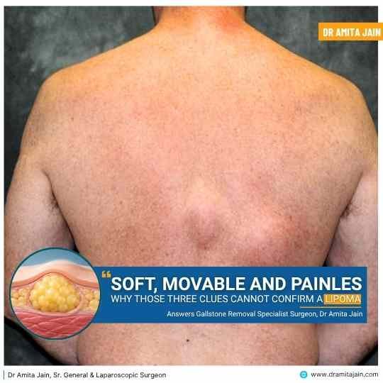
A typical lipoma feels soft or doughy and may move slightly beneath the skin when touched. Most grow slowly and do not cause pain. They commonly appear on the neck, shoulders, back, arms, thighs, and trunk. However, their size, depth, and consistency can vary.
“A lump may feel harmless, but its texture cannot tell the whole story. An accurate diagnosis comes from understanding what lies beneath the skin, not just what the fingers can feel.” Says Dr Amita Jain, specialized surgeon for lipoma in South Delhi.
Why Are These Three Characteristics Not Enough to Confirm a Lipoma?
Softness, mobility, and the absence of pain are not exclusive to lipomas. Other lumps beneath the skin can share these features, making it difficult to identify their exact nature through touch alone.
An epidermoid cyst, for example, may feel rounded and movable. Certain benign soft-tissue growths can also resemble lipomas. Even some serious soft-tissue tumors may initially appear as painless swellings.
A doctor therefore considers several factors together, including the lump’s location, depth, size, consistency, growth pattern, and relationship with surrounding tissues.
What Should You Look for in a Lump?
- Texture: A lipoma often feels soft or doughy, but other growths can have a similar texture.
- Movement: Some lumps move beneath the skin, while others may feel attached to deeper tissues.
- Pain: A painless lump is not necessarily harmless, as some serious growths can develop without discomfort.
- Growth: A lump that gradually increases in size should be medically assessed.
- Location: Whether the lump is superficial or deep beneath the skin can influence the investigations required.
What Other Lumps Can Feel Like a Lipoma?
Several conditions can resemble a lipoma during an initial examination. Their differences may not always be obvious without further assessment.
| Type of Lump | Typical Characteristics | Important Distinction |
| Lipoma | Soft, doughy, usually movable and painless | Made up of mature fat cells and generally grows slowly |
| Epidermoid cyst | Round, often firm or slightly soft, sometimes movable | May have a central opening and can become inflamed or infected |
| Enlarged lymph node | Variable in size, consistency, and mobility | Often associated with infection, inflammation, or other underlying conditions |
| Benign soft-tissue tumour | May be soft or firm depending on the tissue involved | Requires assessment to identify its tissue of origin |
| Soft-tissue sarcoma | May present as a painless, gradually enlarging lump | A malignant growth that requires specialist evaluation |
These descriptions are general patterns, not diagnostic rules. The appearance and feel of a lump alone cannot reliably establish whether it is benign or malignant.

Which Warning Signs Should You Never Ignore?
A lump deserves medical evaluation if it changes in size, becomes increasingly noticeable, or has features that are not typical of a straightforward superficial lipoma.
Pay particular attention to a lump that is growing, measures around 5 cm or more, feels deep beneath the skin, appears attached to underlying structures, or returns after removal. New pain, skin changes, or a noticeable change in consistency also warrant assessment.
A growing or deep lump should be evaluated promptly, even if it is painless. Smaller lumps can also require investigation, so size alone should never determine whether medical advice is necessary.
How Do Doctors Confirm Whether a Lump Is a Lipoma?
Diagnosis usually begins with a clinical examination. The doctor will ask when the lump appeared, whether it has changed in size, and whether it causes discomfort. Its position, depth, consistency, mobility, and relationship with surrounding tissues are also assessed.
Depending on the findings, the following investigations may be recommended:
Clinical Examination: Many typical superficial lipomas can be identified through medical history and physical examination. However, an examination cannot always distinguish a lipoma from other soft-tissue lumps.
Ultrasound: This is commonly used to assess superficial lumps. It helps evaluate their size, depth, composition, and relationship with nearby structures. It may also help distinguish a typical lipoma from a cyst or another type of mass.
MRI: An MRI may be recommended when a lump is deep, large, growing, or has uncertain features on initial assessment. It provides detailed information about soft tissues and helps doctors evaluate the extent and characteristics of the mass.
Biopsy: If the diagnosis remains uncertain or imaging raises concerns, a tissue sample may be required. Examination of the sample under a microscope helps establish the nature of the growth.
Not every suspected lipoma requires all these investigations. The appropriate approach depends on the clinical findings.
Does Every Confirmed Lipoma Need Treatment?
No. Most lipomas are harmless and do not require removal if they are confidently diagnosed, stable, and not causing symptoms.
Treatment may be considered when a lipoma causes pain, interferes with movement, continues to grow, creates cosmetic concerns, or has uncertain diagnostic features. Surgical removal is a common treatment when intervention is necessary.
The decision should depend on the diagnosis, location, size, symptoms, and individual circumstances. Attempting to squeeze, puncture, or remove a lump at home can cause infection, bleeding, and tissue damage.
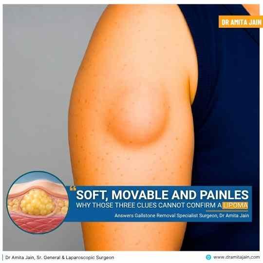
A Reassuring Feel Is Not a Confirmed Diagnosis
A soft, movable, and painless lump may well be a lipoma, but these characteristics are clues rather than proof. Other benign and, less commonly, serious conditions can present in similar ways.
The safest approach is to have a new or changing lump assessed by a qualified doctor rather than relying on how it feels. When necessary, appropriate imaging or tissue examination can help establish the diagnosis and guide treatment.
5 Key FAQs: Understanding Soft, Movable & Painless Lumps
Summary: These three features are common to lipomas, but they are reassuring clues rather than definitive proof.
Answer: While a classic lipoma is composed of mature fat cells that typically feel soft or doughy, move slightly beneath the skin, and cause no pain, several other conditions share these exact physical characteristics. For instance, epidermoid cysts can feel rounded and movable, while certain benign tumors and even some early-stage soft-tissue sarcomas (malignant tumors) can initially present as painless, movable swellings. Because human fingers cannot distinguish the exact microscopic cellular origin through the skin, palpation alone cannot rule out other benign or malignant conditions.
Clinical Key Point: Palpation evaluates surface mechanics, not cellular histology. Touch alone cannot establish benignity.
Summary: Common mimics include epidermoid cysts, enlarged lymph nodes, benign soft-tissue tumors, and soft-tissue sarcomas.
Answer: Multiple growths develop in or near the subcutaneous plane and can be mistaken for a lipoma during an initial self-exam:
• Epidermoid Cyst: A round, slightly soft or firm sac beneath the epidermis, frequently with a subtle central opening (punctum); prone to inflammation.
• Enlarged Lymph Node: A bean-shaped swelling that may be mobile, often reacting to infection, inflammation, or immune triggers.
• Benign Soft-Tissue Tumors: Such as neurofibromas, schwannomas, or fibromas, which can feel soft or rubbery depending on their tissue of origin.
• Soft-Tissue Sarcoma: A rare, aggressive malignant soft-tissue cancer that often starts deceptively as an unassuming, painless, gradually enlarging lump.
Clinical Key Point: Benign cysts and malignant sarcomas can both present as painless masses in their early stages.
Summary: Enlargement, size ≥ 5 cm, depth beneath muscle fascia, fixed anchoring, new pain, or recurrence.
Answer: Any lump that deviates from a small, superficial, stable nodule warrants prompt clinical assessment. Never ignore:
• Progressive Growth: A lump that is visibly enlarging over weeks or months.
• Size ≥ 5 cm: The “golf ball rule”—lumps 5 cm or larger have a higher clinical suspicion index.
• Deep Location / Fixation: Feeling anchored to underlying muscle fascia or bone rather than sliding freely in the subcutaneous fat.
• Changes in Texture: Becoming firmer, harder, or nodular over time.
• New Onset of Pain: Throbbing, tenderness, or compression of adjacent cutaneous nerves.
• Recurrence: A lump reforming in an area where one was previously excised.
Clinical Key Point: A painless lump that is deep, larger than 5 cm, or expanding requires prompt specialist evaluation.
Summary: Through clinical examination, followed by high-frequency ultrasound, MRI for deep or large masses, and biopsy if uncertainty persists.
Answer: Diagnosis follows a structured, evidence-based sequence:
1. Clinical Physical Exam: The doctor inspects the anatomical plane, mobility (including the “slippage sign”), borders, and attachment to adjacent tissues.
2. High-Frequency Ultrasound: The standard first-line imaging tool. It evaluates size, echogenicity (homogeneous hyper-echoic or isoechoic fat vs fluid-filled cyst), vascularity, and depth relative to muscle fascia.
3. Magnetic Resonance Imaging (MRI): Indicated if the mass is deep beneath the fascia, >5 cm, rapidly growing, or ambiguous. MRI differentiates benign adipose tissue from atypical lipomatous tumors or sarcomas.
4. Biopsy & Histopathology: If imaging reveals non-adipose nodules, thick septations, or tissue invasion, a core needle sample is examined under a microscope.
Clinical Key Point: Ultrasound is the first-line imaging tool; MRI is indicated for deep or >5 cm masses; biopsy provides the definitive histology.
Summary: Most confirmed lipomas do not require removal if stable; home squeezing or draining is dangerous and ineffective.
Answer: No, most confirmed lipomas are benign and completely harmless. If a lipoma has been accurately diagnosed, is stable in size, and does not cause pain or functional limitation, standard clinical practice is simple observation without intervention.
Surgical excision or marginal removal is considered if:
• The lump causes pain or impinges on nerves or blood vessels.
• It restricts joint mobility or muscle movement.
• It continues to grow steadily or causes significant cosmetic distress.
• Diagnostic features remain inconclusive and require histopathology.
Crucially, never attempt to squeeze, puncture, lance, or remove a lump at home. Lipomas consist of encapsulated solid fat cells, not liquid pus. Home attempts will not shrink the lump and carry severe risks of bacterial cellulitis, permanent scarring, severe bleeding, and nerve injury.
Clinical Key Point: Asymptomatic lipomas can be left alone. Squeezing or cutting at home creates serious infection and scarring risks.

Dr Amita Jain is one of India’s most distinguished and experienced female surgeons and proctologists, known for her unmatched expertise in general, advanced laparoscopic, and laser proctology surgery. With over 30 years of surgical excellence, Dr Amita Jain has built a reputation for precision, compassionate care, and advanced surgical techniques.
Dr Jain has successfully performed a wide range of complex general surgeries, including both open and minimally invasive procedures, with a strong focus on trauma care, onco-surgical techniques, and reconstructive surgeries. Her medical specialisation includes: gallbladder surgeries, appendix removal surgeries, hernia repair surgeries, Pilonidal Sinus treatments, varicose vein and piles surgeries, fistula surgeries and fissure surgeries.
Dr Jain, currently serves as Senior Consultant and Head of Department (General, Laser, Proctology and Laparoscopic Surgeries) across premier medical networks in South Delhi:
Daffodils by Artemis Hospitals: Spring Meadows, East of Kailash, New Delhi. Book Appointment
Fortis C-DOC Hospital: B-16, Chirag Enclave, New Delhi. Book Appointment
Rosewalk Luxury Multi-Specialty & Maternity Hospital: Panchsheel Park, New Delhi. Book Appointment
Rainbow Children’s Hospital: Malviya Nagar, New Delhi. Book Appointment
Learn More about Dr Amita Jain.
Dr Amita Jain has also authored some of the key medical journals and publications, like ResearchGate, IJSR
Note: This article is written and reviewed by Dr Amita Jain, while referring to her medical journals and publications.